The Digital Portable Ultrasound Scanner has been designed considering the needs of modern health care, providing uninterrupted performance with its rapid image acquisition and high-definition visualization. The robust outer casing and sophisticated temperature control system will make sure that the device continues to work and be trustworthy. Also, the Digital Portable Ultrasound Scanner is a device that aids the long-term data archiving process for efficient medical record management.

In medical imaging, the Digital Portable Ultrasound Scanner is a trustworthy device for different clinical departments. It provides vascular studies support by observing the state of the arteries and veins; in urology, it has a role in the assessment of the bladder and prostate health. The Digital Portable Ultrasound Scanner also enables emergency medical staff to make quick trauma evaluations, directing their actions precisely and efficiently.

The Digital Portable Ultrasound Scanner will proceed to develop as new innovations emerge in artificial intelligence and data analysis. The new models of the Digital Portable Ultrasound Scanner will be able to provide training simulations that experts can use to improve scanning sessions. The increased processing power and connectivity of the Digital Portable Ultrasound Scanner will set new standards of accessibility and accuracy in medical scanning.

In order to extend the service life of the Digital Portable Ultrasound Scanner, it is recommended that users refrain from applying much force during the process of connecting/disconnecting probes. Power cables should always remain dry. The Digital Portable Ultrasound Scanner needs diagnostic tests to ensure that it produces quality images.
Used in hospitals and clinics, the Digital Portable Ultrasound Scanner provides immediate visual feedback for a variety of medical evaluation uses. Converting sound waves into live images, the Digital Portable Ultrasound Scanner allows physicians to easily detect abnormalities. The Digital Portable Ultrasound Scanner assists with making diagnostic processes safer in addition to improving patient outcomes. It possesses an ergonomic shape alongside digital integration capabilities that support simple data sharing and medical record documentation.
Q: What imaging modes are available on the ultrasound scannert? A: It supports multiple modes such as B-mode, M-mode, and color Doppler for diverse diagnostic applications. Q: How does the ultrasound scannert improve diagnostic accuracy? A: By providing high-resolution images and real-time feedback, it enables more precise medical evaluations. Q: Can the ultrasound scannert be used in field or remote settings? A: Yes, its portable versions are designed for mobility and can be used in clinics, hospitals, or mobile healthcare units. Q: What kind of display does the ultrasound scannert use? A: It typically features a high-definition digital display that enhances image visualization and readability. Q: How is data from the ultrasound scannert managed? A: The device allows secure storage, easy access, and export of imaging data through USB or network connections.
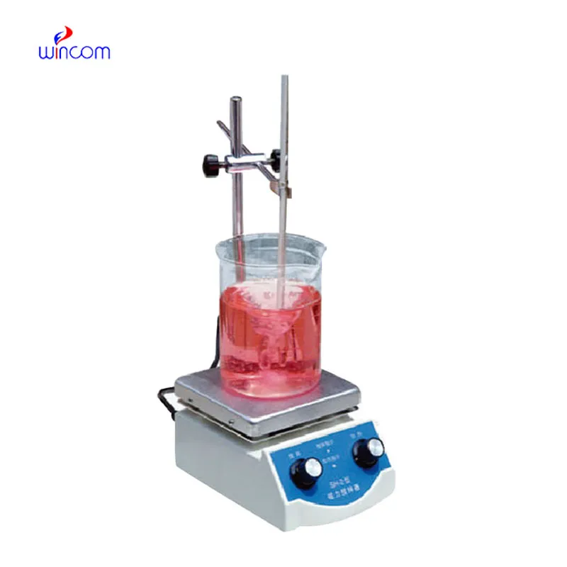
We’ve used this centrifuge for several months now, and it has performed consistently well. The speed control and balance are excellent.
This x-ray machine is reliable and easy to operate. Our technicians appreciate how quickly it processes scans, saving valuable time during busy patient hours.
To protect the privacy of our buyers, only public service email domains like Gmail, Yahoo, and MSN will be displayed. Additionally, only a limited portion of the inquiry content will be shown.
We’re currently sourcing an ultrasound scanner for hospital use. Please send product specification...
I’d like to inquire about your x-ray machine models. Could you provide the technical datasheet, wa...
E-mail: [email protected]
Tel: +86-731-84176622
+86-731-84136655
Address: Rm.1507,Xinsancheng Plaza. No.58, Renmin Road(E),Changsha,Hunan,China

 af
af
 es
es
 ar
ar
 tr
tr
 sw
sw
 pt
pt
 th
th
 ur
ur
 bn
bn
 ne
ne
 vi
vi
 km
km
 lo
lo
 de
de
 ru
ru
 fi
fi
 nl
nl
 fa
fa
 fr
fr
 ko
ko