The body tube microscope combines technological innovation and practical design, with distortion-free, clear-quality imaging at every magnification. The mechanical stability and focus precision controls of the body tube microscope ensure accurate specimen positioning. The body tube microscope enhances sample visibility in varying light conditions using a strong illumination system. Optional camera adapters and measuring software are offered to extend its use, making it suitable for various scientific and educational environments.

Applications of the body tube microscope cross into different spheres. It enables disease diagnosis by examining tissue sample and blood smears in medicine. In materials science, the body tube microscope is employed to examine crystal structures, coatings, and composites. In life sciences research, it is used in visualization of cell morphology, patterns of growth, and intracellular action. The body tube microscope also offers quality inspection for production with precision in semiconductor fabrication and microfabrication. It is used in museums and conservation laboratories to examine pigments and fibers in artifacts from ancient times.

The body tube microscope will also evolve by being combined with new quantum and digital technologies. Greater processing speed and improved imaging will capture microscopic motion in real time. Artificial intelligence will decipher complex biological and material structures more accurately than ever before. The body tube microscope will likely consist of interchangeable modular components that can be replaced or reconfigured based on specific research needs. The body tube microscope will remain vital as the scientific frontiers continue to push the frontiers of the unexplored in nature.

In the interest of precision and reliability, the body tube microscope should be constantly exposed to cleanliness and maintenance. Switch it off at all times before adjusting or cleaning parts. The lenses may be cleaned with alcohol-free cleaners lightly to avoid scratching. Rotary components such as knobs and stage mechanisms value light lubrication at regular intervals. The body tube microscope must be stored away from direct sunlight and vibration. Professional checking once a year ensures optical alignment is not affected and prevents wear from invisible damage.
A body tube microscope is a convenient tool that magnifies microscopic materials that are invisible to the naked eye. It allows researchers, scientists, and students to view cells, microorganisms, and sensitive materials with careful attention at microscopic sizes. Modern body tube microscope models combine optical precision with electronic technology to give high-definition images and fine focusing. They are widely applied in biology, medicine, and material sciences for research, study, and instruction. With high-performance lenses and illumination systems, a body tube microscope enhances visualization to enable users to examine texture, shape, and structure at the microscopic level with utmost clarity and accuracy.
Q: What is a microscope used for? A: A microscope is used to magnify tiny objects or structures, allowing detailed observation of cells, microorganisms, and materials that are invisible to the naked eye. Q: How often should a microscope be calibrated? A: To maintain measurement accuracy and ensure accurate focus during research or analysis, regular calibration should be performed, typically once or twice a year. Q: What type of light source is commonly used in a microscope? A: Most modern microscopes use LED or halogen light sources, which provide stable light and adjustable brightness for clear images at a wide range of magnifications. Q: Can a microscope be connected to a computer? A: Yes, many microscope models feature USB or HDMI ports that allow image capture and digital display through specialized imaging software. Q: How should a microscope be stored when not in use? A: A microscope should be covered with a dust shield and stored in a cool, dry location to prevent contamination and protect optical components from humidity.
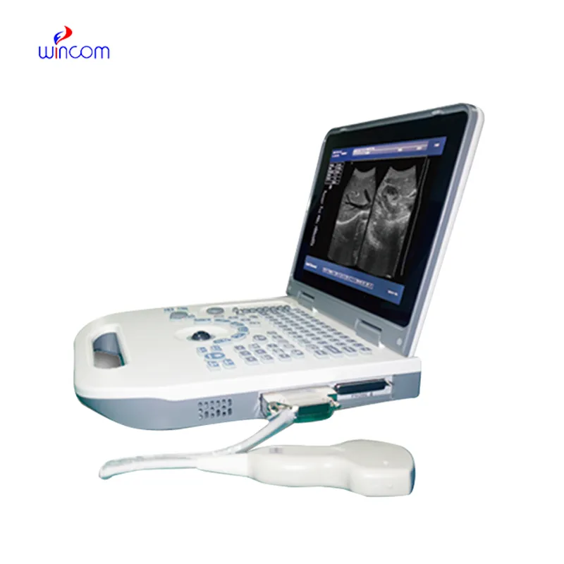
This ultrasound scanner has truly improved our workflow. The image resolution and portability make it a great addition to our clinic.
The delivery bed is well-designed and reliable. Our staff finds it simple to operate, and patients feel comfortable using it.
To protect the privacy of our buyers, only public service email domains like Gmail, Yahoo, and MSN will be displayed. Additionally, only a limited portion of the inquiry content will be shown.
We’re currently sourcing an ultrasound scanner for hospital use. Please send product specification...
Could you share the specifications and price for your hospital bed models? We’re looking for adjus...
E-mail: [email protected]
Tel: +86-731-84176622
+86-731-84136655
Address: Rm.1507,Xinsancheng Plaza. No.58, Renmin Road(E),Changsha,Hunan,China

 af
af
 es
es
 ar
ar
 tr
tr
 sw
sw
 pt
pt
 th
th
 ur
ur
 bn
bn
 ne
ne
 vi
vi
 km
km
 lo
lo
 de
de
 ru
ru
 fi
fi
 nl
nl
 fa
fa
 fr
fr
 ko
ko