In hospital and research facility settings, digital baby cradling scale provides the critical mass measurement that is needed for delicate analyses. It is the case that reagents, samples, and medicines are weighed with the highest level of precision. Laboratory staff regard the digital baby cradling scale as their helper in making the measurements, carrying out calibrations, and performing quality assurance. Besides being of great assistance in the above activities and ensuring accurate measurement in clinical diagnosis, experimental research, and drug response monitoring, digital baby cradling scale also enhances overall laboratory performance and has a positive impact on the dependability of analytical results.

In pathology laboratories, digital baby cradling scale finds its usage during the staining compounds preparation and the tissue processing additives application. The proper mass measurement guarantees the same composition of the reagent, and this, in turn, affects the performance of the stain and the interpretation under the microscope. This application helps to maintain the same standards in pathology workflows and minimizes the differences between test batches. By using strict preparation conditions, digital baby cradling scale plays a role in the stability of diagnosis in hospital pathology departments.
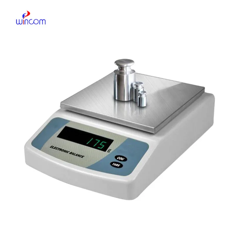
At the medical institutions that are research-driven, digital baby cradling scale will change to facilitate the analytical methods with higher sensitivity that are in the pipe. The future might bring along the possibility of ultra-low mass samples being accurately measured in molecular diagnostics and sophisticated drug research. This turning development will not only enlarge the experimental capacities of hospital-based research labs but also open new fronts in medical innovation through analytics.
In order to keep digital baby cradling scale in a good condition consistent calibration practices are needed that follow hospital laboratory protocols. Scheduled calibration checks are performed to maintain the reliability of measurements during daily activities involving analysis. Conditions in the environment such as temperature and the amount of air that moves around should be kept under control so as to prevent drift. The people operating the machines should make sure that there are no sudden changes in load and that the weighing pan is not subjected to excessive force. Through adhering to controlled handling practices, digital baby cradling scale is always trusted for pharmaceutical preparation and medical research activities.
digital baby cradling scale is of great importance for developing and validating laboratory methods. One of the most important prerequisites for getting correct analytical results is the weighing of reference standards, buffers, and chemicals with absolute accuracy. Laboratory personnel depend on digital baby cradling scale for constant concentration reproduction and hence, reliable testing. Its superb sensitivity plus reliable performance indeed make it a primary instrument for validating methods in the clinical, hospital, and pharmaceutical lab settings.
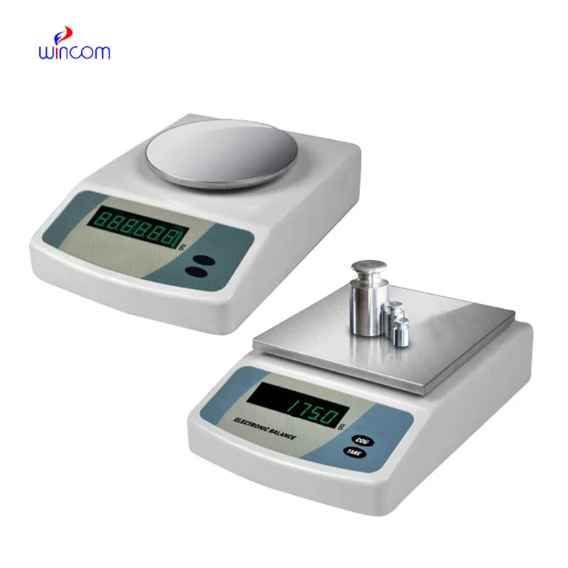
Q: What is the main purpose of an Analytical Balance? A: Its purpose is mainly to measure very tiny sample masses with the utmost precision in laboratories and hospitals. Q: What is the typical weighing range of an Analytical Balance? A: The weighing range for the majority of analytical balances is from 0 up to some grams with a resolution of micrograms or milligrams. Q: What environmental controls are necessary for an Analytical Balance's operation? A: Airflow, vibration, and temperature changes should not only be avoided but also prevented in the room where the scale is situated. Q: Is an Analytical Balance permitted in a hospital laboratory? A: Yes, it has indeed found widespread usage for the preparation of reagents, calibra¬tion, and drug development applications. Q: What should be the frequency of calibration for an Analytical Balance? A: The calibration interval is subject to the degree of use and the particular laboratory requirements.
This ultrasound scanner has truly improved our workflow. The image resolution and portability make it a great addition to our clinic.
This x-ray machine is reliable and easy to operate. Our technicians appreciate how quickly it processes scans, saving valuable time during busy patient hours.
To protect the privacy of our buyers, only public service email domains like Gmail, Yahoo, and MSN will be displayed. Additionally, only a limited portion of the inquiry content will be shown.
We are planning to upgrade our imaging department and would like more information on your mri machin...
We’re looking for a reliable centrifuge for clinical testing. Can you share the technical specific...
E-mail: [email protected]
Tel: +86-731-84176622
+86-731-84136655
Address: Rm.1507,Xinsancheng Plaza. No.58, Renmin Road(E),Changsha,Hunan,China

 af
af
 es
es
 ar
ar
 tr
tr
 sw
sw
 pt
pt
 th
th
 ur
ur
 bn
bn
 ne
ne
 vi
vi
 km
km
 lo
lo
 de
de
 ru
ru
 fi
fi
 nl
nl
 fa
fa
 fr
fr
 ko
ko