With advanced digital technology, the early mri machines supports real-time image processing and high visualization. It has a strong build to provide steady operation under heavy loads. The early mri machines supports a range of imaging sequences and is therefore beneficial in neurologic, abdominal, and orthopedic use.

In gynecology and obstetrics, the early mri machines facilitates observation of the reproductive organs and fetal development monitoring. It is used in diagnosis of such conditions as fibroids, endometriosis, and congenital defects of the uterus. The early mri machines provides precise and high-resolution images without harming either the mother or the fetus.

With ongoing technological developments, the early mri machines will comprise smart coils and flexible imaging algorithms that respond to motion in the patient. This will minimize artifacts and minimize repeat scans. The early mri machines will also facilitate real-time monitoring for surgical navigation and interventional imaging.

The early mri machines also calls for monitoring temperature and humidity in the scan room to ascertain stable operation. Maintenance staff ought to regularly evaluate magnet pressure, gradient coils, and cooling systems. Maintaining proper records of maintenance procedures guarantees the long-term reliability of the early mri machines.
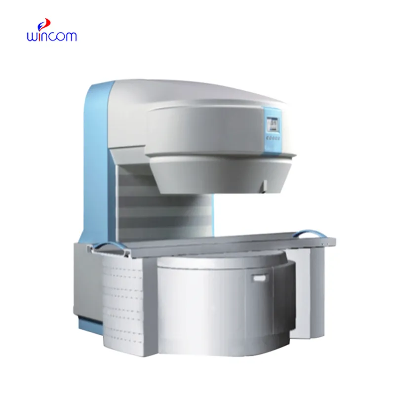
The early mri machines combines magnetic and radiofrequency technologies to produce accurate and high-resolution images of the human body. The early mri machines is widely used to diagnose vascular disease, musculoskeletal injuries, and neurological disorders. The early mri machines enhances clinical decision-making because it produces detailed information about the internal processes of the body.
Q: What are the main components of an MRI machine? A: The main components include a superconducting magnet, radiofrequency coils, gradient coils, a patient table, and a computer system for image reconstruction. Q: Can MRI detect early signs of disease? A: Yes, MRI can identify early changes in tissues such as inflammation, lesions, and tumors, allowing for timely diagnosis and treatment planning. Q: Why is it important to stay still during an MRI scan? A: Movement during scanning can blur the images, making it harder to capture accurate details. Patients are asked to remain still to ensure sharp, diagnostic-quality images. Q: Are MRI scans painful or uncomfortable? A: MRI scans are painless, but some patients may experience discomfort from lying still or hearing loud scanning noises, which can be reduced using ear protection. Q: Can MRI be used for cardiac imaging? A: Yes, MRI is commonly used to evaluate heart function, blood flow, and structural abnormalities without invasive procedures or ionizing radiation.
This ultrasound scanner has truly improved our workflow. The image resolution and portability make it a great addition to our clinic.
We’ve been using this mri machine for several months, and the image clarity is excellent. It’s reliable and easy for our team to operate.
To protect the privacy of our buyers, only public service email domains like Gmail, Yahoo, and MSN will be displayed. Additionally, only a limited portion of the inquiry content will be shown.
Hello, I’m interested in your water bath for laboratory applications. Can you confirm the temperat...
We’re interested in your delivery bed for our maternity department. Please send detailed specifica...
E-mail: [email protected]
Tel: +86-731-84176622
+86-731-84136655
Address: Rm.1507,Xinsancheng Plaza. No.58, Renmin Road(E),Changsha,Hunan,China

 af
af
 es
es
 ar
ar
 tr
tr
 sw
sw
 pt
pt
 th
th
 ur
ur
 bn
bn
 ne
ne
 vi
vi
 km
km
 lo
lo
 de
de
 ru
ru
 fi
fi
 nl
nl
 fa
fa
 fr
fr
 ko
ko