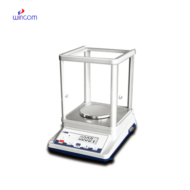
In hospital and clinical laboratories, electronic benefit transfer delaware balance supplies exact mass measurements that are very important to experimental integrity. It allows the very accurate weighing of the reagents, chemicals, and components of the patient's sample. Technicians in the laboratory use electronic benefit transfer delaware balance for the maintenance of reproducibility, validation of method, and monitoring of therapy. electronic benefit transfer delaware balance assures the consistency of analytical data, so, it increases the efficiency of the hospital laboratory, makes the research more reliable, and through accurate measurement of the materials in the laboratory, it becomes part of the patient care that is of high quality.
In microbiology labs, electronic benefit transfer delaware balance is utilized for the preparation of culture media and analytical additives. Accurate weighing guarantees that the nutrient composition for microbial growth and testing is consistent. This application helps to produce reliable cultures, conduct antimicrobial studies, and do infection research. By keeping the mass control precise, electronic benefit transfer delaware balance supports reproducibility in microbiological workflows in clinical laboratories.
The era of electronic benefit transfer delaware balance in hospitals will go beyond the traditional settings and embrace multidisciplinary research environments. As the partnership of clinical, pharmaceutical, and biomedical teams becomes more robust, the analytical balances will cater to different experimental needs. By taking on various analytical actions, electronic benefit transfer delaware balance will still be a fundamental tool in contemporary hospital laboratory ecosystems.
For electronic benefit transfer delaware balance to last long, professionals must do scheduled servicing as per the laboratory guidelines. Internal cleaning and checking of parts will help to slow down and eventually stop the performance degradation. Hospitals or labs that maintain structured service intervals not only enjoy lesser downtimes but also higher accuracy, thus they can carry on with seamless analytical operations.
electronic benefit transfer delaware balance plays an important role in the hospital pharmacy in the accurate formulation of medications, intravenous solutions, and compounded drugs. Even slight alterations in the weight of drugs can change the effectiveness of the drug and endanger the patient's safety. The Pharmacy Technicians rely on electronic benefit transfer delaware balance for the correct dosing and checking of the active ingredients. The tool's accuracy guarantees the dependable preparation of drugs, the observance of the rules, and the quality control of the overall hospital pharmacy activities.
Q: What is the impact of temperature on the performance of analytical balance? A: The changes in temperature can lead to drift and weighing inconsistency. Q: Are analytical balances the only ones used in research laboratories? A: They are very important also for other processes such as sample preparation and improving the accuracy of the experiment. Q: How long does it usually take for an analytical balance to warm up? A: Warm-up times differ from one model to another, but an adequate stabilizing period increases the reliability of the measurement. Q: Is it possible for analytical balances to save weighing data? A: Internal memory or external data transfer are the two ways in which many models can achieve this feature. Q: Would it be necessary to undergo training if one wants to operate an analytical balance? A: Basic laboratory training will be enough to make sure that the balance is being used correctly.
This ultrasound scanner has truly improved our workflow. The image resolution and portability make it a great addition to our clinic.
The microscope delivers incredibly sharp images and precise focusing. It’s perfect for both professional lab work and educational use.
To protect the privacy of our buyers, only public service email domains like Gmail, Yahoo, and MSN will be displayed. Additionally, only a limited portion of the inquiry content will be shown.
I’d like to inquire about your x-ray machine models. Could you provide the technical datasheet, wa...
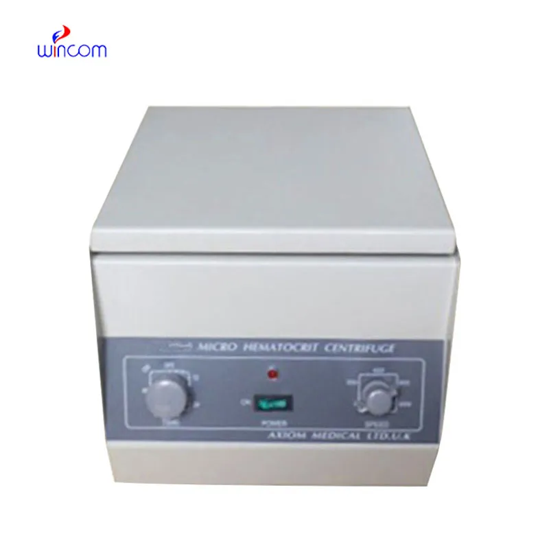
We’re looking for a reliable centrifuge for clinical testing. Can you share the technical specific...
E-mail: [email protected]
Tel: +86-731-84176622
+86-731-84136655
Address: Rm.1507,Xinsancheng Plaza. No.58, Renmin Road(E),Changsha,Hunan,China

 af
af
 es
es
 ar
ar
 tr
tr
 sw
sw
 pt
pt
 th
th
 ur
ur
 bn
bn
 ne
ne
 vi
vi
 km
km
 lo
lo
 de
de
 ru
ru
 fi
fi
 nl
nl
 fa
fa
 fr
fr
 ko
ko