The ge ultrasound machine works by integrating cutting-edge image optimization features that reduce artifacts and improve detail recognition thereby increasing diagnostic accuracy. It caters to multilingual users and sets for personal usability requirements for global needs. The device provides imaging of the same quality regardless of the patient type or clinical condition.

The ge ultrasound machine is a very significant diagnosis tool used in obstetrics for fetal monitoring and pregnancy development detection. It indirectly affects the cardiology field by providing information about the hearts and blood flow dynamics. Furthermore, the ge ultrasound machine is very important in diagnosing abdominal problems, especially issues with the liver, kidneys, and gallbladder. Still, it is also being used in musculoskeletal diagnoses for spotting ligament and tendon injuries.

The future of the ge ultrasound machine may include enhancements based on artificial intelligence algorithms that can improve images and offer automatic measurements. Better transducer technologies may offer higher resolution and increased sensitivity. The ge ultrasound machine may have an increasing role to play in personalized medicine because it may offer continuous monitoring capabilities.

Care of the ge ultrasound machine involves much more than cleanup. Environmental monitoring and mechanical protection are also part of the process. The ge ultrasound machine should not be subject to either vibration or shock. The ge ultrasound machine should be backed up periodically to retain vital images.
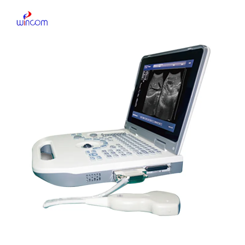
The ge ultrasound machine represents an advanced form of medical imaging technology that transforms sound waves into high-definition visual data. It is widely used for evaluating organ health, tracking fetal development, and detecting vascular conditions. The ge ultrasound machine ensures real-time monitoring and fast diagnostic results, supporting effective clinical workflows.
Q: What is the primary function of an ultrasound scannert? A: Ultrasound scanners are designed to create real-time images of internal organs, tissues, and blood flow using high-frequency sound waves. Q: How does the ultrasound scannert ensure clear imaging results? A:It uses advanced converter technology and digital processing to enhance image clarity and contrast. Q: In what medical fields is the ultrasound scannert commonly used? A: It is widely used in obstetrics, cardiology, urology, radiology, and emergency medicine. Q: Is the ultrasound scannert safe for repeated use? A: Yes, it is non-invasive and does not emit radiation, making it safe for frequent diagnostic applications. Q: Can the ultrasound scannert store and share imaging data? A: Yes, it supports data storage, retrieval, and digital transfer for easy integration with hospital systems.
This ultrasound scanner has truly improved our workflow. The image resolution and portability make it a great addition to our clinic.
The centrifuge operates quietly and efficiently. It’s compact but surprisingly powerful, making it perfect for daily lab use.
To protect the privacy of our buyers, only public service email domains like Gmail, Yahoo, and MSN will be displayed. Additionally, only a limited portion of the inquiry content will be shown.
Hello, I’m interested in your centrifuge models for laboratory use. Could you please send me more ...
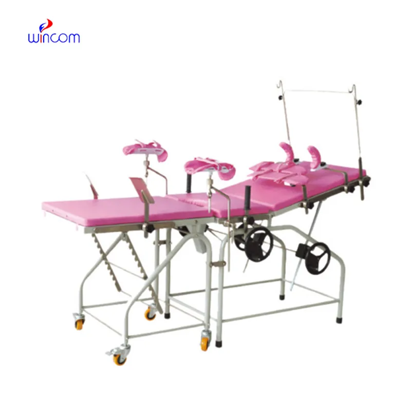
We’re interested in your delivery bed for our maternity department. Please send detailed specifica...
E-mail: [email protected]
Tel: +86-731-84176622
+86-731-84136655
Address: Rm.1507,Xinsancheng Plaza. No.58, Renmin Road(E),Changsha,Hunan,China

 af
af
 es
es
 ar
ar
 tr
tr
 sw
sw
 pt
pt
 th
th
 ur
ur
 bn
bn
 ne
ne
 vi
vi
 km
km
 lo
lo
 de
de
 ru
ru
 fi
fi
 nl
nl
 fa
fa
 fr
fr
 ko
ko