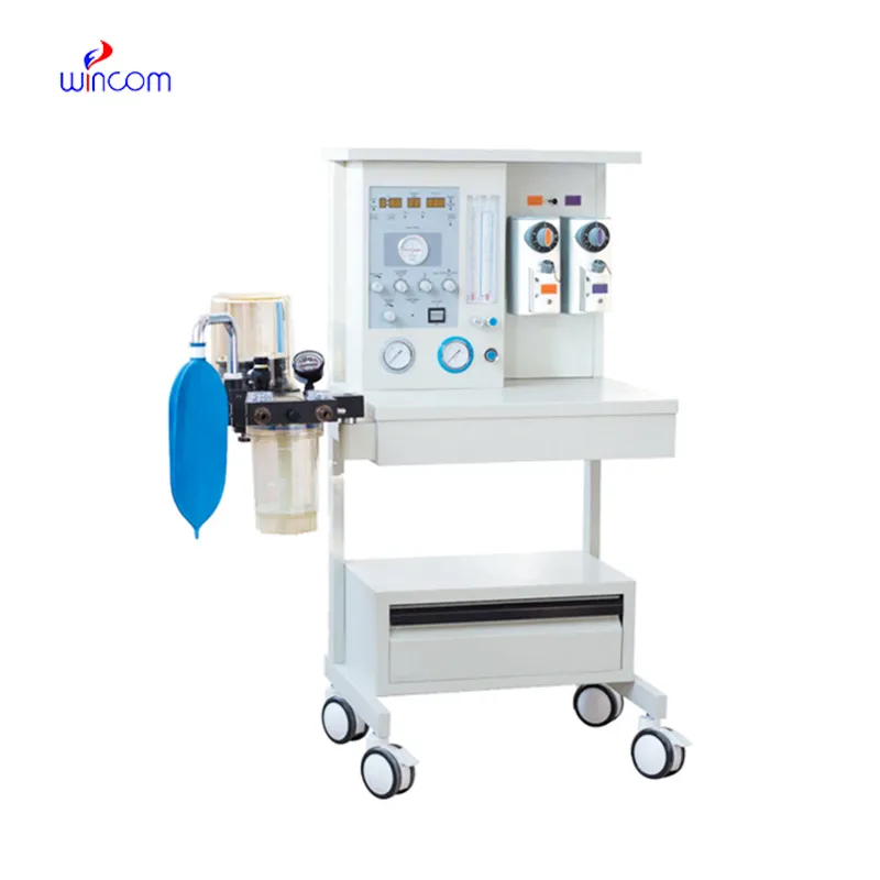
mri anesthesia machine is capable of perfecting the anesthetic gas mixtures and at the same time providing mechanical ventilation for the patients during the surgical process. Its monitoring systems keep a check on the various vital parameters like oxygen saturation, airway pressure, and tidal volume. This device is widely in use in hospitals and clinical laboratories for the exact purpose of keeping sedation and respiratory function under control. The collaboration of alarm systems, flow controls that can be modified, and a real-time display permits anesthesiologists to react without delay to physiological changes. mri anesthesia machine offers safe and effective anesthesia delivery which is why it is considered a must-have in operating rooms, emergency care units, and research places.

In mri anesthesia machine, the training tool is used in teaching hospitals for anesthesia education purposes. Medical students and residents get to learn anesthesia methods by looking at gas delivery systems, watching different ventilation modes, and monitoring indicators. The device provides both simulated and supervised clinical practice, and it lets the trainees grasp the real patient responses in the controlled conditions. So, by combining practical training with constant monitoring, mri anesthesia machine not only helps to the professional growth in the hospital education system but also enhances the clinical skills in anesthesia management area.
In the future, mri anesthesia machine will very probably add the features of ventilation that are more automated and suited to patients' breathing habits. Smart algorithms could help the doctors by changing the settings of ventilation according to the constant feedback. This development could lessen the need for human control in long operations without losing the accuracy of respiratory control. The same consistency in anesthesia delivery could be achieved in hospitals and laboratories doing research. These advancements are characteristic of the new medical environments where the anesthesia equipment will be more adaptive and responsive.
mri anesthesia machine are the most important things in hospital maintenance and cleaning procedures. The external surfaces need to be disinfected with approved disinfectants to totally avoid the transfer of germs. For example, the internal parts like vaporizers need to be checked regularly to make sure that the residue does not harm the operation. In the labs and operating rooms, highly strict cleanliness rules are applied which are beneficial to both patients and medical personnel. Moreover, proper drying after cleaning saves the equipment from moisture damage. These practices of care and maintenance ensure the safe use and keep the mri anesthesia machine constant in all the medical departments performance.
In contemporary medical centers, mri anesthesia machine is implemented for the purpose of ensuring stability in anesthesia throughout intricate surgical procedures. It manages the amount of anesthetic gasses and at the same time gives continuous feedback on the patient's breathing and oxygen levels. The apparatus permits uninterrupted observation of life signs and makes it possible to carry out modifications according to the needs of the patient. Moreover, mri anesthesia machine facilitates accurate anesthesia management, thereby mitigating the dangers linked to both over- and under-sedation and leading to the best possible patient outcomes in all hospital operating rooms and clinical labs.
Q: How frequently should Anesthesia Machine be checked? A: The machine should be routine checked before daily use and also during the scheduled maintenance cycles. Q: Is the Anesthesia Machine movable within a hospital? A: There are lots of models available that are specifically designed with wheels that allow easy movement between the departments. Q: Can it be connected with hospital monitoring systems? A: There are some models that are designed for the connection with central monitoring and data systems. Q: What is the procedure when the oxygen supply is cut off? A: The safety mechanisms will alert the clinicians and will also help in keeping the patient protected. Q: Is the use of this equipment limited to specially trained personnel only? A: Definitely, only properly trained personnel will be able to use the machine safely and efficiently.
The hospital bed is well-designed and very practical. Patients find it comfortable, and nurses appreciate how simple it is to operate.
The delivery bed is well-designed and reliable. Our staff finds it simple to operate, and patients feel comfortable using it.
To protect the privacy of our buyers, only public service email domains like Gmail, Yahoo, and MSN will be displayed. Additionally, only a limited portion of the inquiry content will be shown.
I’d like to inquire about your x-ray machine models. Could you provide the technical datasheet, wa...
We’re currently sourcing an ultrasound scanner for hospital use. Please send product specification...
E-mail: [email protected]
Tel: +86-731-84176622
+86-731-84136655
Address: Rm.1507,Xinsancheng Plaza. No.58, Renmin Road(E),Changsha,Hunan,China

 af
af
 es
es
 ar
ar
 tr
tr
 sw
sw
 pt
pt
 th
th
 ur
ur
 bn
bn
 ne
ne
 vi
vi
 km
km
 lo
lo
 de
de
 ru
ru
 fi
fi
 nl
nl
 fa
fa
 fr
fr
 ko
ko