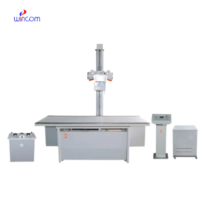
The x ray wall scanner has been designed keeping in mind the needs of modern healthcare. The system comes equipped with capabilities such as real-time imaging adjustments and intelligent exposure. The digital detector provides high uniformity of images, thus supporting accurate interpretations. The x ray wall scanner comes with connectivity solutions that ensure smooth integration with digital radiology networks.

The x ray wall scanner is commonly used in medical imaging to examine skeletal trauma, lung disease, and dental anatomy. The x ray wall scanner assists physicians in diagnosis of fractures, infection, and degenerative disease. The x ray wall scanner is also used in orthopedic surgery intraoperatively. In emergency medicine, it provides rapid diagnostic information that allows clinicians to assess trauma and internal injury rapidly.

Future versions of the x ray wall scanner will combine energy-efficient technology with high-resolution imaging. Predictive analytics integration will enable early disease detection and personalized screening. Global telemedicine networks will also be enabled by the x ray wall scanner, extending access to diagnosis in underserved populations.
The x ray wall scanner needs regular maintenance to function at its best. Technicians need to regularly inspect exposure controls, cooling systems, and image sensors. The x ray wall scanner has to be run within prescribed usage boundaries, and annual recalibration needs to be planned to maintain radiation accuracy as well as uniform imaging quality.
In today's healthcare system, the x ray wall scanner continues to be an integral part of diagnostic imaging. The x ray wall scanner provides precise visual data that helps in disease detection and assessment of an injury. The x ray wall scanner has digital sensors and the capability to improve images. The x ray wall scanner helps in quick and effective medical imaging.
Q: What makes an x-ray machine different from a CT scanner? A: An x-ray machine captures a single 2D image, while a CT scanner takes multiple x-rays from different angles to create 3D cross-sectional views. Q: How is image quality measured in an x-ray machine? A: Image quality depends on factors like contrast, resolution, and exposure settings, which are adjusted based on the target area being examined. Q: What power supply does an x-ray machine require? A: Most x-ray machines operate on high-voltage power systems, typically between 40 to 150 kilovolts, depending on their intended use. Q: Can x-ray machines be used for dental imaging? A: Yes, specialized dental x-ray machines provide detailed images of teeth, jaws, and surrounding structures to support oral health assessments. Q: How does digital imaging improve x-ray efficiency? A: Digital systems allow instant image preview, faster diagnosis, and reduced need for retakes, improving workflow efficiency in clinical environments.
We’ve used this centrifuge for several months now, and it has performed consistently well. The speed control and balance are excellent.
The centrifuge operates quietly and efficiently. It’s compact but surprisingly powerful, making it perfect for daily lab use.
To protect the privacy of our buyers, only public service email domains like Gmail, Yahoo, and MSN will be displayed. Additionally, only a limited portion of the inquiry content will be shown.
Hello, I’m interested in your water bath for laboratory applications. Can you confirm the temperat...
We’re looking for a reliable centrifuge for clinical testing. Can you share the technical specific...
E-mail: [email protected]
Tel: +86-731-84176622
+86-731-84136655
Address: Rm.1507,Xinsancheng Plaza. No.58, Renmin Road(E),Changsha,Hunan,China

 af
af
 es
es
 ar
ar
 tr
tr
 sw
sw
 pt
pt
 th
th
 ur
ur
 bn
bn
 ne
ne
 vi
vi
 km
km
 lo
lo
 de
de
 ru
ru
 fi
fi
 nl
nl
 fa
fa
 fr
fr
 ko
ko